(work in progress)
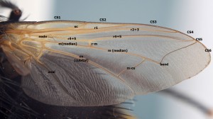
Wing venation is a very important part of keying any tachinid so here are a few pointers to help you. Let's start by familiarising ourselves with the names of each wing vein:
Questions that you will come across in the keys:
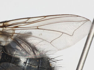
- Does the wing have a petiole? This is often described in other ways, such as whether wing cell R5 is open or closed or whether the median vein meets r4+5 before the wing margin … but essentially it is the same test. The median vein runs out towards the wing edge and then (in most tachinids) bends forward. It then usually just meets the wing edge just below the point where vein r4+5 also touches the wing edge, but occasionally it meets r4+5 earlier, creating a short stalk or petiole that continues to the wing edge. Here are some examples of wings without petioles:
and here are some examples of wings with strong petioles:
Occasionally the median vein meets r4+5 at the wing margin (so not petiolate but also not open either) but the key couplets should allow for this.
[insert photos of examples meeting at the wing margin – some Eriothrix etc.]
- Is vein m-cu at approximately 45-degrees to the cubital vein? This one should be easy with practice and it is a great way to quickly spot Voriini in any sample (Voria, Athrycia, Cyrtophleba etc.). If you haven’t seen this feature before though it can be a bit tricky but the secret is not to ponder too hard – if it is strongly angled then you will know it – if you are struggling to work out whether it is 45-degrees or more like 60-degrees then it probably isn’t angled. Here are some examples:
[insert photos of Voria wings]
- Does the median vein “disappear”? On some species (eg. Actia lamia & Ocytata pallipes) the median vein disappears or peters out before it reaches the wing margin. This is a rare feature and is usually easy to see:
- Does the median vein have a “shadow fold”? In some tachinids when you look at the median vein, where it bends, you will see a short appendix that extends from the bend a little but in some Exoristini this has been reduced to a vague crease in the wing membrane. Although it can be difficult to see sometimes it is a very useful feature. Remember that what you are looking for isn't a vein - it really is just a slight crease in the wing membrane. Here is an example of a shadow fold in Exorista tubulosa:
- Is the second wing edge segment hairy beneath? This one was very confusing in Belshaw because he used the wrong terminology to explain it, calling it the sub-costal vein when it is actually just the second segment of the costa (CS2 in my diagram). The test is only used to split off Phryxe heraclei from the other Phryxe spp. but it is a feature that you will check often because Phryxe spp. are so common. First turn the wing so that you are looking at the ventral surface and find the sub-costal vein and vein r1 - the costal section that we are interested runs between them. Every tachinid has tiny bristlets along the leading edge of the whole wing but most will have a bare area of vein between these bristlets and the wing membrane. Phryxe heraclei on the other hand will have between 1-5 tiny bristles on this bare area of vein but they might be very hard to spot and it is worth checking both wings if you can.
[insert a photo of a Phryxe heraclei wing & a Phryxe vulgaris wing]
- Do the hairs on r4+5 extend to the cross vein r-m? This test is something you will do on nearly every tachinid and you soon get used to it but on your first few you might get a bit worried about how precisely to judge the hairiness.
[insert photos of an Athrycia wing, an Actia wing & an Exorista wing]


